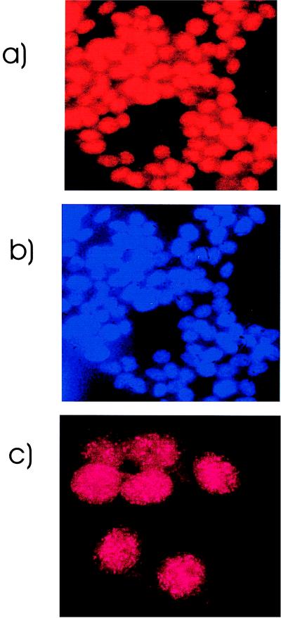
Figure 5.
Immunofluorescence analysis of SCP-1 expression. Indirect immunofluorescence with SCP-1 antiserum (a) and counterstaining with 4′,6-diamidino-2-phenylindole (b) reveals a nuclear staining with a punctated staining pattern (c) in Cheng cells that scored positive for SCP-1 in RT–PCR and in Western blot analysis. This pattern was observed in almost all interphase nuclei, indicating that SCP-1 expression in tumor cells is not restricted to a particular cell cycle phase as observed in sperm cells.

