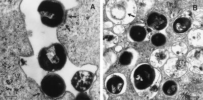
Figure 1.
DC internalize S. gordonii via conventional phagocytosis. DC were incubated with bacteria for different time periods at a ratio of 10 bacteria to 1 DC, then washed and processed for transmission electron microscopy. (A) Thirty minutes after infection, bacteria were contacting the cell membrane and inducing local thickening of plasma membrane (arrows) (magnification, ×39,000; bar represents 3.9 μm). (B) Four hours after infection, bacteria were found in phagolysosomes in a partially degraded form (arrow) at a magnification of ×28,700; bar = 2.9 μm.

