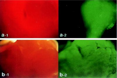
Figure 1.
Rat hearts were transduced with Ad.EGFP by using either the catheter-based technique (b) or direct injection into the left ventricular wall (a). Forty-eight hours following delivery of adenovirus encoding for EGFP, the left ventricles of the hearts were removed and visualized with white light (a-1 and b-1) and at 510 nm with single excitation peak at 490 nm of blue light (a-2 and b-2). As shown in b-2, the expression pattern observed after catheter delivery is grossly homogeneous. In contrast, the expression pattern is localized after direct injection as shown in a-2. Of note, with the direct injection, the surrounding tissue exhibits no background fluorescence (a-2).

