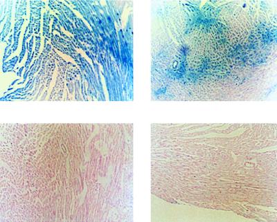
Figure 2.
Expression of β-gal in left ventricular sections 2 days following infection with Ad.βgal and Ad.PL. (Upper) Photomicrographs of two left ventricular sections stained for β-gal 2 days following infection with Ad.βgal. These sections show the variability of β-gal expression within the same heart with the catheter-based method of gene delivery. (Lower) Photomicrographs of two left ventricular sections stained for β-gal 2 days following infection with Ad.PL. No β-gal expression is observed.

