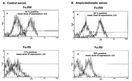
Figure 9.
Flow cytometric analysis of FcγRIII and FcɛRII surface expression in rabbit ASM cells. Cells treated for 24 hr with either 10% control serum or 10% atopic asthmatic serum were stained with FITC-conjugated human mAbs specific for the low-affinity FcγRIII (A) and FcɛRII (CD23) (B) receptors. Activated B-cells (8.1.6) were used as a positive control for the CD23 receptor. The level of nonspecific background staining was measured in both the control- and atopic asthmatic- serum-treated cells by staining with FITC-conjugated isotype control antibodies. Note that the rabbit ASM cells express surface protein for both FcγRIII and FcɛRII receptors. In contrast to FcγRIII receptor expression, which is unaltered in the presence of atopic asthmatic serum, expression of the FcɛRII receptor is increased by >2-fold (i.e., from 17 to 36% in the presence of atopic asthmatic serum.

