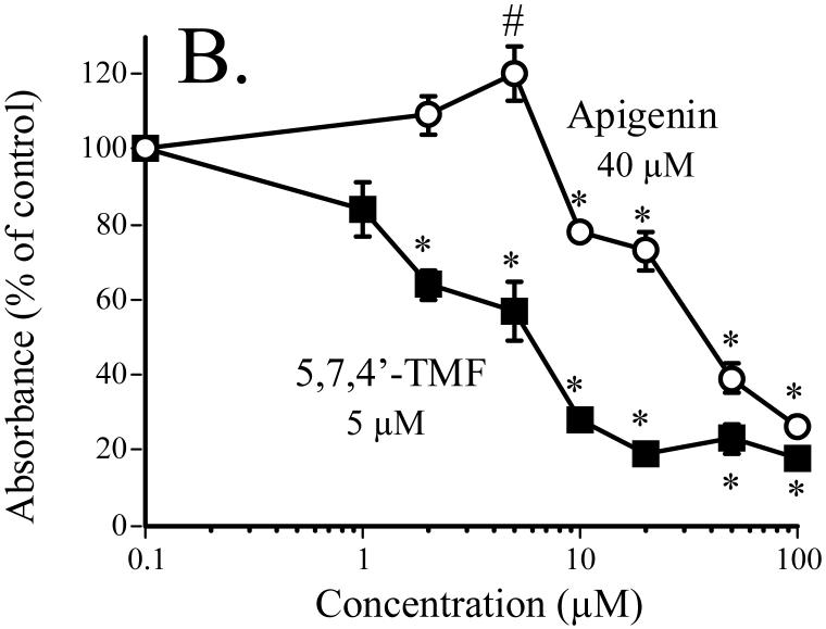
Fig. 4.
Effect of 5,7,4′-TMF compared to the unmethylated analog apigenin on SCC-9 cell proliferation. Cell proliferation, expressed as percent of control (DMSO-treatment), was measured as BrdU incorporation into cellular DNA after a 24-h exposure of the cells to the flavones [31]. The numbers shown in the figure are the calculated IC50 values. From [31].
* significantly lower than control, P < 0.05. # significantly higher than control, P < 0.05.

