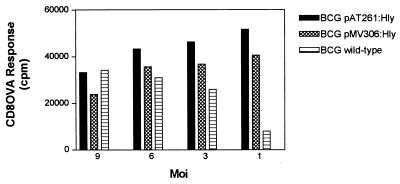
Figure 6.
Presentation of OVA to CD8 T cells by MHC class I molecules of macrophages infected with BCG pAT261:Hly, BCG pMV306:Hly, or BCG wild-type microorganisms. Interferon-γ-activated and BM A3.1A7 cells (H-2Kb haplotype) were incubated with BCG pAT261:Hly (solid bars), BCG pMV306:Hly (cross-hatched bars), or BCG wild-type (horizontal hatched bars) bacteria in presence of OVA (50 μg/ml) at the indicated moi for 24 h, fixed with 1% paraformaldehyde, washed, and subsequently cocultured with OVA257–264-specific CD8 T–T hybridoma cells RF33.70 for 24 h. Supernatants were assayed for IL-2. Data are presented in triplicate from a representative experiment; the SEM never exceeded 10%. Noninfected BM A3.1A7 macrophages with or without OVA coculture never exceeded 4,000 cpm. Experiments were performed three times with similar results.

