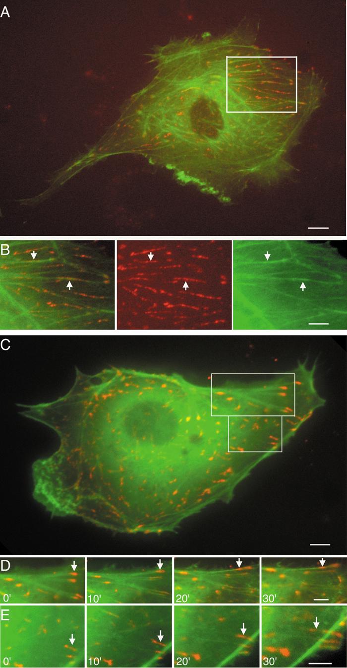
Fig. 2. Dynamic motion of paxillin-fibers and paxillin-clusters in association with actin filaments in living ECs co-transfected with DsRed2-paxillin (red) and GFP-actin (green).

(A) A static EC. (B row) Boxed area in A (Movie S4B series). Arrows point to the dynamic motion of paxillin-fibers on SFs. (C) An EC subjected to shear stress. (D and E rows) Boxed areas in C. Paxillin-clusters (arrows) show disassembling and sliding on actin SFs. Superimposition of DsRed2-paxillin and GFP-actin images appears as yellow (Movie S4DE series). Scale bars: A, C = 10 μm; B, D, E = 8 μm.
