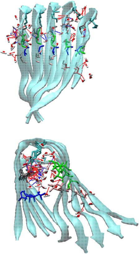
FIGURE 10.
Interior hydration of Aβ models. Top panel is side view perpendicular to fibril axis, whereas bottom panel is top view along fibril axis. Broken red lines indicate H-bonds between water molecules and side chains of inward-pointing residues, whereas broken blue lines indicate H-bonds between penetrated water molecules.

