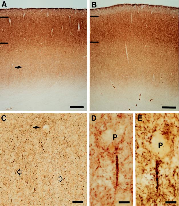
Figure 1.
Laminar distribution and overall intensity of GAT-1 immunoreactivity in area 46 is similar in matched normal control (A) and schizophrenic (B) subjects (see Table 1, triad 3). Hash marks indicate the layer 1–2 and 3–4 borders. At higher magnification (C), GAT-1-labeled axon cartridges (open arrows) are readily identified amid the diffuse punctate immunoreactivity. Solid arrow indicates the same blood vessel in A and C. Individual GAT-1-immunoreactive axon cartridges are located below the unlabeled cell bodies of pyramidal neurons (P) in normal control (D) and schizophrenic (E) subjects. [Bars = 500 μm (A and B), 50 μm (C), and 10 μm (D and E).]

