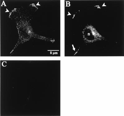
Figure 5.
Confocal microscopy of immunostained Kv1.4-transfected MDCK-F cells. (A) Lowermost scan section. Only the nuclear periphery was imaged in this section. Kv1.4 was strictly localized at the leading edge of the lamellipodium (arrowheads). No accumulation of the Kv1.4 protein could be detected in plasma membrane surrounding the cell body. (B) Upper section of the same cell as in A (approximately 400 nm above). Kv1.4 was clearly detected at the leading-edge membrane (arrowheads), as in A. The intracellular staining was restricted to the endoplasmic reticulum and/or to the Golgi network surrounding the nucleus. The fluorescently labeled cell extension is marked with an arrow. This staining is most likely because of unspecific binding of the secondary antibody. (C) Mock-transfected MDCK-F cells showed no signal.

