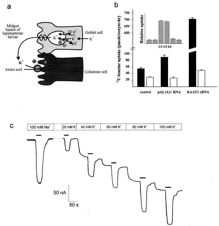
Figure 1.
(a) Schematic model of the midgut epithelium of lepidopteran larvae showing the relationship between KAAT1 and other apical membrane components. The apical membrane of goblet cells contains an H+ vacuolar type ATPase in parallel with a K+/2H+ antiporter that secretes K+ into the lumen while maintaining a transapical voltage of ≈−270 mV. The voltage, attenuated to ≈−240 mV, appears across the apical (brush border) membrane of electrically coupled columnar cells where it drives K+-coupled amino acid transport from lumen to cell via KAAT1. (b) Na+-coupled uptake of 200 μM 14C-leucine (1 h) in Xenopus oocytes injected with mRNA prepared from M. sexta larval midgut or with cRNA synthesized by in vitro transcription using KAAT1 cDNA. Open bars indicate water injection. The black bar in the control group indicates injection of total RNA. (Inset) Uptake of 200 μM 14C-leucine (1 h) in oocytes injected with size-fractionated poly(A)+ RNA from M. sexta larval midgut. All data points represent the mean ± SEM from 6–8 oocytes. (c) Leucine (1 mM) evoked uptake currents mediated by KAAT1. A representative oocyte expressing KAAT1 was voltage-clamped at −50 mV and superfused with uptake solution containing 100 mM Na+ or specified concentrations of K+. When K+ was being tested, Na+ was replaced with choline+. Leucine was applied where indicated by black bars.

