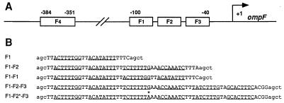
Figure 1.
The ompF regulatory region. (A) The locations of the two separate OmpR binding regions upstream of the start point of ompF transcription (arrow marked +1) and the locations of individual OmpR binding sites (F1, F2, F3, F4) within these regions are indicated. (B) The sequences of the nontemplate strands for the five double-stranded oligonucleotides used in the DNA migration retardation assays are indicated. The DNA fragments consist of two parts: DNA sequences from the ompF regulatory region (uppercase) and flanking sequences that are not present at ompF (lowercase). The underlined bases indicate the positions of the half-sites in the individual sites (6). The asterisk indicates the position of the G-to-A substitution in the mutant F2 site.

