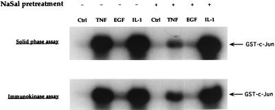
Figure 1.
Inhibition of TNF-induced JNK activation by NaSal. Serum-starved FS-4 cells (19) were treated for 1 h with 20 mM NaSal. They were then either left untreated (Ctrl) or treated for 15 min with TNF (20 ng/ml), EGF (30 ng/ml), or IL-1-α (4 ng/ml). Lysates were generated, and used directly in a protein kinase assay with GST-c-Jun (solid-phase assay, Upper). An antibody to JNK1 and protein G-agarose were used to form an immunoprecipitate from the same lysates that then was used in the kinase assay with GST-c-Jun (immunokinase assay, Lower). Phosphorylation reactions were initiated by the addition of [γ-32P]ATP and terminated by the addition of protein sample buffer. The reaction mixtures were separated by SDS/PAGE, and phosphorylated GST-c-Jun protein was visualized by autoradiography.

