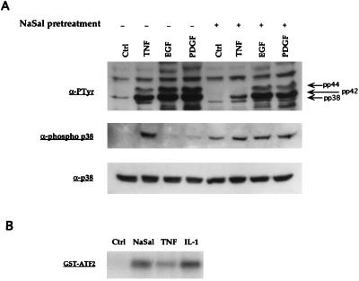
Figure 3.
Activation of p38 MAPK by NaSal. (A) Western blot analyses of lysates generated from FS-4 cells. Serum-starved cells were treated for 1 h with 20 mM NaSal. They were then either left untreated (Ctrl) or treated for 15 min with TNF (20 ng/ml), EGF (30 ng/ml), or PDGFbb (50 ng/ml). Lysates were blotted with antibodies against phosphotyrosine (α-PTyr, Top), antibodies specific for the tyrosine-phosphorylated form of p38 MAPK (Middle), or antibodies to p38 MAPK protein (Bottom). Arrows in Top denote positions of the phosphorylated MAPKs pp38, pp42, and pp44. (B) Assay of p38 kinase activity. COS-1 cells were transfected with an epitope-tagged p38 MAPK expression vector (pCMV-Flag-p38 MAPK; ref. 8), serum-starved for 16 h, and then left untreated (Ctrl) or stimulated for 15 min with NaSal (20 mM), TNF (20 ng/ml), or IL-1 (4 ng/ml). Flag-p38 MAPK was then immunoprecipitated, and kinase activity was assayed by incubating the immunoprecipitates with [γ-32P]ATP and GST-ATF2 as substrate. The reaction mixtures were separated by SDS/PAGE, and phosphorylated GST-ATF2 was visualized by autoradiography. Equal expression of Flag-p38 MAPK in each lysate prior to immunoprecipitation was confirmed by Western blot analysis (data not shown).

