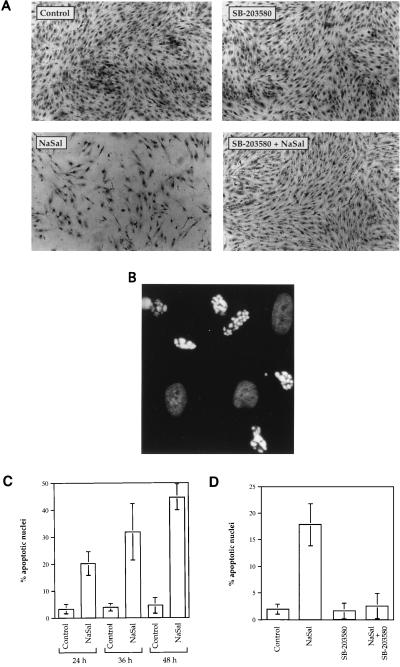
Figure 4.
Induction of apoptosis by NaSal and its inhibition by SB-203580. (A) Cytotoxicity of NaSal in FS-4 cultures is inhibited by SB-203580. Serum-starved cells were either left untreated (Control), or treated for 48 h with SB-203580 (10 μM), NaSal (20 mM), or a combination of SB-203580 and NaSal at these same concentrations. For the combination treatment, cells were incubated with SB-203580 for 1.5 h before NaSal addition. Shown are photomicrographs of cells that were fixed and stained with naphthol-blue-black (23). (B) NaSal induces cell death by apoptosis. FS-4 cells were treated for 12 h with 20 mM NaSal, stained with Hoechst dye 33342, and examined by fluorescence microscopy. Photomicrograph demonstrates typical appearance of brightly staining highly vesiculated apoptotic nuclei with condensed chromatin. Also visible are less intensely stained nonapoptotic oval nuclei. (C) Time course of the induction of apoptosis by NaSal. Serum-starved FS-4 cells were either left untreated (Control) or treated with 20 mM NaSal for the indicated time periods. Hoechst dye-stained nuclei from at least six different microscopic fields were counted. Data shown are means ± SD from a representative experiment. Similar results were obtained in three other experiments. (D) Inhibition of NaSal-induced apoptosis by SB-203580. Serum-starved FS-4 cells were either left untreated (Control) or treated for 36 h with SB-203580 (10 μM), NaSal (20 mM), or a combination of SB-203580 and NaSal. For the combination treatment, cells were incubated with SB-203580 for 1.5 h before NaSal addition. Hoechst dye-stained nuclei from at least six different microscopic fields were counted. Data shown are means ± SD from a representative experiment. Similar results were obtained in three other experiments.

