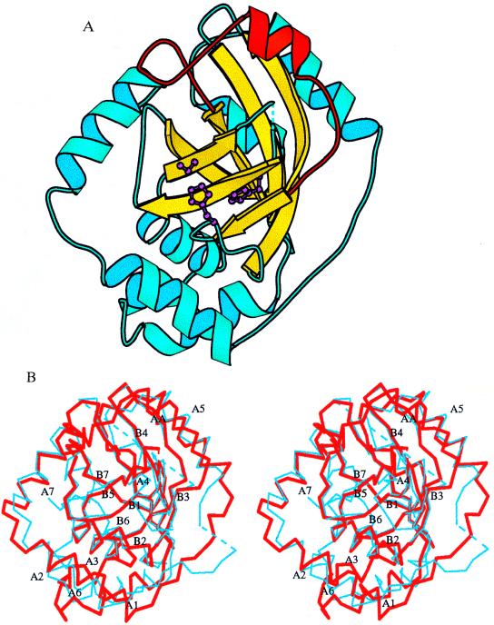
Figure 2.
The monomeric structure of the VZV protease. (A) The core β-barrel (yellow), the catalytic triad (purple), and the AA loop (red). (B) Stereoview of the superposition between the VZV (thick red lines) and CMV (thin blue lines) protease structures. The secondary elements of the VZV protease are labeled. The diagram was drawn with the program molscript (19).

