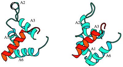
Figure 3.
(Left) The dimer interface of VZV protease. (Right) CMV protease. Helix A6 of one of the monomers is shown in red; A1, A2, A3, and A6 of the other are shown in blue. The two A6 helices are not parallel to each other in VZV protease. The segments containing helices A2 assume quite different conformations in VZV and CMV proteases.

