Figure 4.
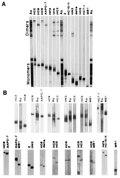
Separation and analysis of crosslinked proteasome subunit pairs. (A) Separation of crosslinked proteasome subunit pairs. Proteasomes were crosslinked with sulfo-EGS and analyzed by SDS/PAGE and immunoblotting. Lanes Au were stained for total protein with colloidal gold (Aurodye; Amersham). Other lanes were incubated with subunit specific antibodies as indicated. (B) Individual subunit dimers were excised from one- or two-dimensional gels and the crosslinkers cleaved. The subunits from the dimers were then identified by a new round of SDS/PAGE and immunoblotting with all 14 subunit-specific antibodies. The subunits from individual crosslinked dimers are shown pairwise. For each dimer the remaining 12 antibodies showed no specific reaction. For dimers isolated only from one-dimensional PAGE, the lanes marked Au shows parallel lanes stained for total protein with colloidal gold to prove absence of other subunits.

