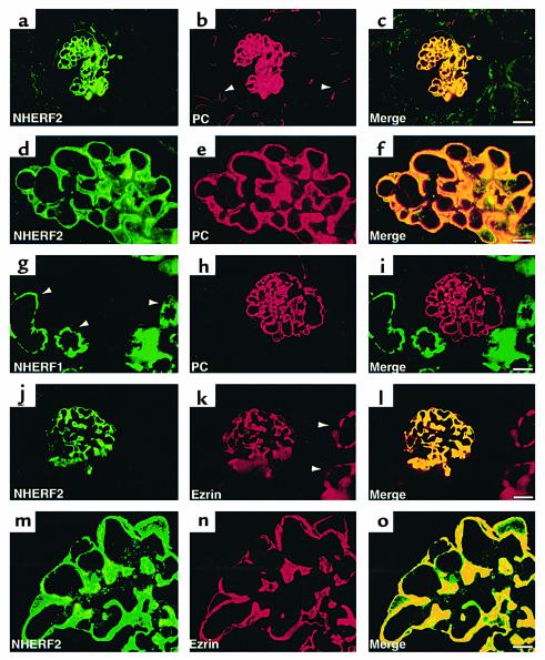
Figure 4.
Localization of NHERF1, NHERF2, PC, and ezrin in the normal rat kidney. Strong staining for both NHERF2 (a and d) and PC (b and e) is seen in GECs in the glomerulus. PC is also present in the endothelium of peritubular capillaries (b, arrowhead) where NHERF2 is not seen. The yellow signal in the merged images (c and f) demonstrate overlap in the staining of GECs. In contrast, NHERF1 (g) is expressed mainly in the apical region of proximal tubules (arrowheads) and is not seen in glomeruli where PC is present (h and i). NHERF2 (j and m) also colocalizes with ezrin (k and n) in GECs where their staining overlaps (yellow staining in l and o). Ezrin is also expressed in proximal tubules (k, arrowheads) where NHERF1 (g) but not NHERF2 (l) is detected. Rat kidneys were processed for semithin (0.5 μm) cryosectioning, followed by double staining with mAbs anti-PC (5A) or anti-ezrin (3C12) and polyclonal anti-NHERF1 or anti-NHERF2, as described in Methods. Bars: (a–c and g–l) 20 μm; (d–f and m–o) 5 μm.

