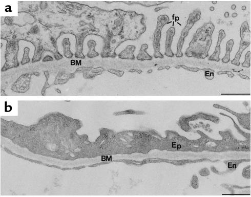
Figure 7.
Morphologic alterations seen in PAN-treated kidney. (a) Portion of a glomerular capillary from a normal, untreated rat showing the typical organization of the foot processes (fp) of the glomerular epithelium. (b) Portion of a capillary from a PAN nephrotic rat showing disruption of the foot process organization of GECs (Ep) and loss of the filtration slits between foot processes. BM, basement membrane; En, endothelium. Bar, 0.5 μm.

