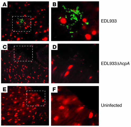
Figure 8. Detection of HCP on thin sections of infected pig intestinal explants.
After infection with the wild-type EDL933 (A) or the EDL933ΔhcpA (C), the tissues were sliced and immunostained with anti-HCP antibody. The DNA of pig intestinal cells and the bacteria are stained in red (with propidium iodide) while HCP are stained in green. Note the absence of HCP in the hcpA mutant. Mock-infected tissues (E) were used as negative controls. B, D, and F are enlarged images of A, C, and E, respectively.

