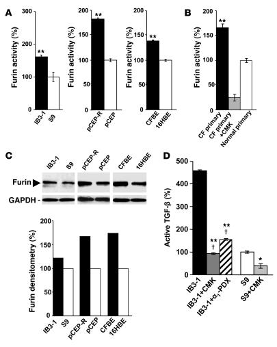
Figure 1. Furin levels are elevated in CF cells.
Furin activity was measured by monitoring cleavage of fluorogenic substrate boc-RVRR-amc in extracts from lung epithelial cells. One unit of activity was defined as the amount of enzyme required to liberate 1 pmol of AMC from boc-RVRR-amc. (A) IB3-1, pCEP-R, and CFBE (all 3 cell lines with a CF phenotype) and S9, pCEP, and 16HBE (all 3 cell lines with CFTR-corrected or non-CF phenotype). (B) Furin activity in primary human (CF and normal) lung epithelial cells measured in the presence or absence of the furin inhibitor CMK. (C) Furin western blot and densitometry analysis. (D) Human lung epithelial cells after treatment with furin inhibitors CMK and α1-PDX. Production of TGF-β was measured in IB3-1 (CF) and S9 (CFTR corrected) epithelial cells using TGF-β–responsive luciferase assay. CMK, 50 μM; α1-PDX, 5 μM. Data are presented as percentage of furin or TGF-β, for which 100% corresponds to protein levels in non-CF cells (n = 3 experiments). Mean ± SEM (*P < 0.05, **P < 0.01; †P ≥ 0.05).

