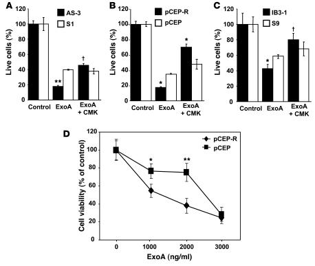
Figure 4. CF cells are more sensitive to P. aeruginosa ExoA.
CF and matched non-CF respiratory epithelial cells were cultured overnight in 96-well plates with media containing 10% serum. Cells were incubated with ExoA for 24 hours. Cell viability was measured by adding WST-1 as described in Methods. (A) AS1 normal human respiratory epithelial cells stably transfected with CFTR antisense construct (inducing CF phenotype) and their matching control S1 (cells stably transfected with the sense construct). (B) pCEP-R (overexpressing the dominant-negative CFTR R domain) and pCEP (mock-transfected). (C) IB3-1 and S9 (CFTR corrected). Human lung epithelial cells were incubated with furin inhibitor CMK (50 μM) for 24 hours at 37°C. (D) Dose-dependent cytotoxicity of ExoA. Lung epithelial cells were treated for 24 hours with increasing concentrations of P. aeruginosa ExoA (0 to 3,000 ng/ml). Data are presented as percentage of live cells, for which 100% corresponds to ExoA untreated cells (n = 3 experiments). Mean ± SEM (*P < 0.05, **P < 0.01, †P ≥ 0.05).

