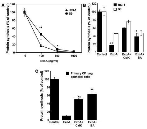
Figure 5. CF cells show increased sensitivity to ExoA inhibition of protein synthesis.
CF and non-CF cells were incubated in media containing either increasing concentrations of ExoA for 24 hours (A) or 100 ng ExoA/ml (B) with IB3-1 and S9 (CFTR-corrected) cells. (C) CF primary lung epithelial cells in the presence or absence of furin inhibitor CMK (50 μM) or treated with 100 nM bafilomycin A1 (BA). Protein synthesis levels were determined by measuring the incorporation of [3H] leucine into TCA-precipitable cellular proteins and are expressed as percentage of control cells that received no toxin. Mean ± SEM for 3 separate experiments; *P < 0.05, **P < 0.01, †P ≥ 0.05.

