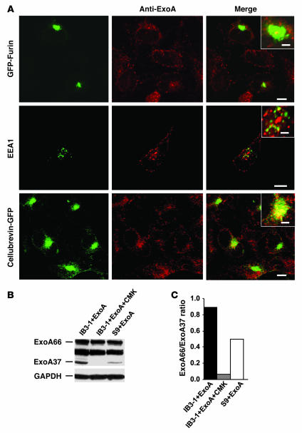
Figure 6. Furin-dependent processing of ExoA is increased in CF cells.
(A) Localization of ExoA in CFTR mutant IB3-1 cells by fluorescence microscopy (insets, close apposition). Cells were transfected with GFP-furin or cellubrevin-pHluorin GFP and incubated with ExoA conjugated to secondary Alexa Fluor 568 (red). EEA1 was visualized using primary human anti-EEA1 antibody and Alexa Fluor 468–conjugated antibody (green). Scale bars: 5 μm; 2 μm (insets). (B) SDS-PAGE analysis of ExoA processing in IB3-1 (CFTR mutant) cells, IB3-1 cells incubated with CMK (50 μM), and S9 (CFTR-corrected IB3-1) cells. Proteins in cell extracts were separated by SDS-PAGE and immunoblotted with anti-ExoA. ExoA66, full size, inactive ExoA toxin precursor; ExoA37, proteolytically processed 37-kDa active ExoA fragment. (C) Densitometry analysis of ExoA66/ExoA37 ratio.

