
Figure 1. Temporospatial distribution of type XXVII collagen in the primary ossification center.
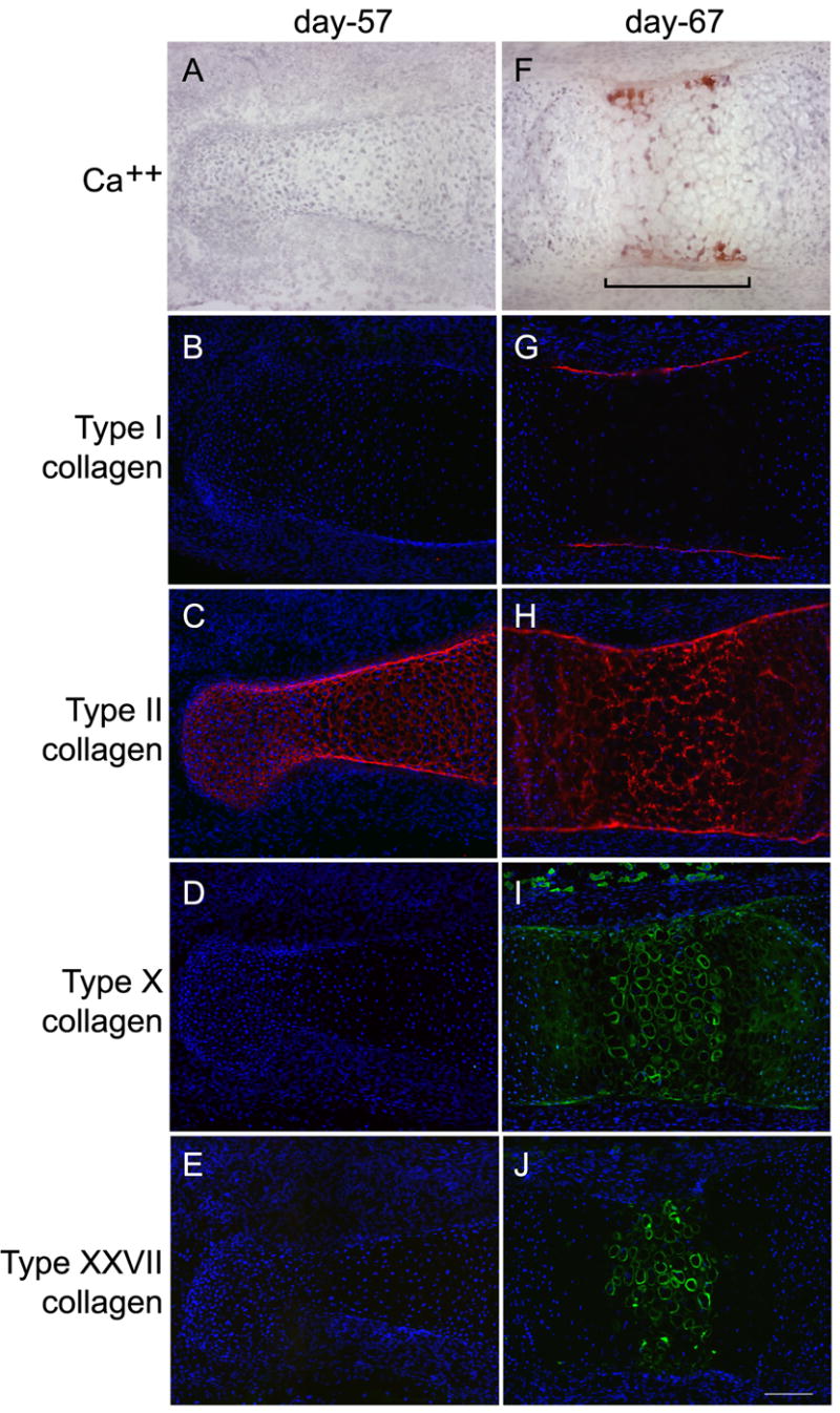
Cartilaginous primordial middle phalanges from 57- and 67-day human fetal fingers were sectioned and stained with alizarin red, which binds calcium (A, F), and with antibodies directed against collagen types I (B, G), II (C, H), X (D, I), and XXVII (E, J). At day 57, there was no cartilage calcification and, of the four collagens, only type II (the predominant cartilage collagen) was present. At day 67, cells at the primary ossification center (bracket) were hypertrophic and the matrix surrounding these cells was calcified. Type I collagen (the main protein of bone) was detected only in the perichondrium. Type II collagen remained throughout the structure. Type X collagen was prominent in the ECM surrounding hypertrophic cells. Type XXVII collagen was detected in the ECM around hypertrophic cells and its pattern of distribution overlapped, in part, with that of calcification and type X collagen. Magnification bar is 125μM.