
Figure 2. Localization of type XXVII collagen in the growth plate.
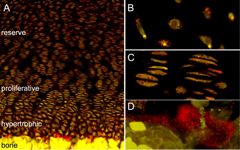
Tissue from a 20-week-old human femoral growth plate was sectioned and interrogated with an antibody directed against type XXVII collagen (red). Type XXVII collagen was detected in the three cartilaginous zones: hypertrophic, proliferative, and reserve. However, staining was most intense in the hypertrophic region, at the interface of newly deposited bone and cartilage (A). Higher magnification of reserve (B) and proliferative (C) zones showed type XXVII collagen on the cell surfaces. Increased magnification of the hypertrophic zone at the cartilage-bone boundary showed type XXVII collagen surrounding chondrocytes (D).