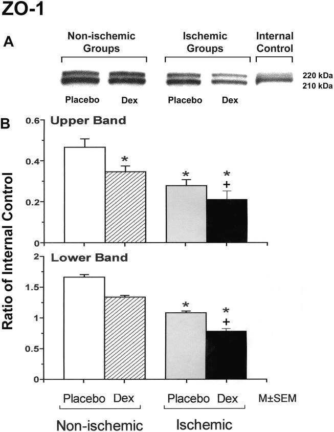
Figure 4.
Representative Western immunoblots and bar graphs of the upper and lower protein bands of ZO-1 expression in the cerebral cortex of the fetuses of placebo (Placebo) and dexamethasone (Dex) treated ewes in the non-ischemic groups on the left, and ischemic groups on the right. ZO-1 is expressed as two isoforms as a result of alternative RNA splicing differing in the presence of an 80-amino acid region referred to as "motif-a": the 235 kDa upper band corresponds to the ZO-1α+ isoform, and the 225 kDa lower band corresponds to the ZO-1α- isoform. The expression patterns are dynamic and cell specific during development [24,26]. Bar legends as for figure 1. Open bars n=6, hatched bars n=6, gray bars n=6 and solid bars n=5 for the upper band and n=6 for the lower band. There were no statistical differences in upper/lower band ratios among the experimental groups (Data not shown).*P<0.05 versus the to placebo/control group. +P<0.05 versus the placebo/ischemic group.

