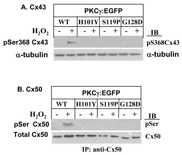
Figure 4. Lack of phosphorylation of Cx43 on Ser368 or Cx50 on serines by PKCγ C1B mutants.
A. Stably-transfected cells were treated with 100 μM H2O2 for 20 min. Phosphorylation of Cx43 on Ser368 was determined by Western blot using anti-phosphoSer368 Cx43 antibodies. α-tubulin levels were shown as controls. B. Cx50 was immunoprecipitated by anti-Cx50 antibodies. The immunoprecipitates were resolved by SDS-PAGE and immunoblotted with anti-phospho-serines antibodies (i.e., site of phosphorylation not presently identified). Cx50 levels were shown as controls.

