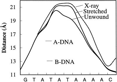
Figure 2.
The 3′–3′ phosphorus distances (Å) for successive dinucleotide base pairs along the TATA box sequence for the x-ray structure of the TATA box (1) and the simulated stretched and unwound conformations (the curve shown is a polynomial fit to the successive P–P distances). The values for canonical A- and B-DNA are shown for comparison.

