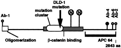
Figure 1.
Antibodies recognize different epitopes of the APC protein. Schematic representation of the APC protein as adapted from (12). The mutation cluster region is indicated by the oval. β-catenin-binding region is indicated by a shaded rectangle. □, Oligomerization region; ○, mAbs that recognize distinct APC epitopes used in the present study. Antibody Ab-4, whose epitope has not been mapped precisely, was made against a 300-amino acid peptide beginning at • and proceeding to the end of the APC protein. The solid line represents the APC region that was used to produce the polyclonal rabbit sera APC64. The arrow marks the point of APC protein truncation in the colon cancer cell line DLD-1. The circled Ns·· indicate two potential nuclear localization sequences beginning at amino acids 1773 and 2054.

