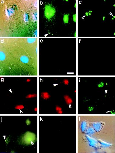
Figure 2.
Localization of APC protein in 184A1 cells using immunofluorescence microscopy. 184A1 cells were grown on glass slides prior to fixation and immunofluorescence microscopy using Ab-4, an antibody specific for APC protein (b and c) or using Ab-4 preincubated with an APC peptide (e). b and c are photographs of the same group of cells taken at two focal distances to more clearly capture cell edge staining (b, solid arrowhead) and nuclear staining (c, open arrowhead). APC protein appears in a punctate pattern throughout the cytoplasm with areas of protein concentration at one edge (solid arrowhead). In addition, APC protein appears throughout the nuclei with a few areas of concentration (c). a and d are DIC and DAPI views of the fluorescence views shown in b, c, and e, respectively. Controls include: f, 184A1 cells stained with nonspecific antibody IgG1; g–i, 184A1 cells stained for APC using antibodies Ab-2 (g), Ab-6 (h) or APC64 (i); j, APC staining of T47D cells. For each antibody, both edge staining (solid arrowhead) and nuclear staining (open arrowhead) are apparent. In k, DLD-1 cells that express only truncated APC protein were stained using the C terminal antibody Ab-4 to demonstrate staining specificity. l is the corresponding DIC and DAPI view of the cells shown in k. (Bar = 10 μm.)

