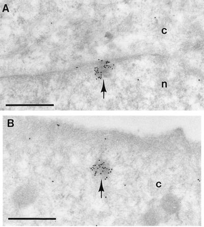
Figure 2.
Localization of Ro protein in cultured keratinocytes by immunogold labeling with antiserum C. Gold particles (10 nm) accumulate over electron-opaque bodies (arrows) at the nuclear border (A) and in the cytoplasm (B). c, Cytoplasm. Glutaraldehyde fixation was 1.6%. Image produced by uranyl acetate staining. (Bars = 0.5 μm.)

