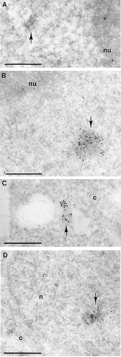
Figure 3.
Colocalization of hY RNA and Ro protein. Gold particles, 5 nm in diameter, label the hY RNA, whereas the 10-nm gold particles label the Ro protein. Formaldehyde fixation was 4%. Image produced by uranyl acetate staining. (A) Part of a nucleus of a HeLa cell. A cluster of 5-nm gold particles only (arrow) is observed within the nucleoplasm. Individual 5-nm and 10-nm gold particles are present. nu, nucleolus. (B) Part of a nucleus of a HEp-2 cell. A cluster of 10-nm gold particles only (arrow) labels an electron-opaque spot within the nucleoplasm in which individual 10-nm gold particles are scattered. nu, Nucleolus. (C) HEp-2 cell. A cluster of mixed 5-nm and 10-nm gold particles (arrow) is present in the cytoplasm (c). (D) HEp-2 cell. A cluster of mixed 5-nm and 10-nm gold particles (arrow) is present within the nucleoplasm over an electron-opaque spot, which appears as highly contrasted fibrils. c, Cytoplasm; n, nucleus. (Bars = 0.5 μm.)

