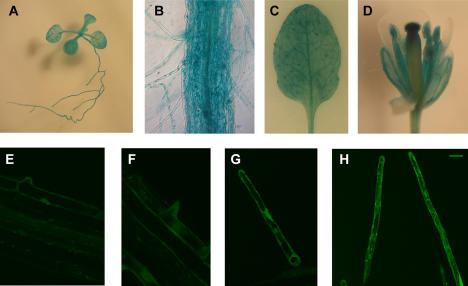
Figure 3. MRH2 promoter activity in root hairs.
Tissue/cell type expression patterns were revealed by histochemical examination of the 955 bp MRH2 promoter activity using the GUS (A–D) or GFP (E–G) reporters. Shown are a 2-week-old young seedling (A), part of the root with root hairs (B), a leaf (C) in which trichomes were strongly stained but epidermal cells were weakly stained, and a flower (D). Root hairs at various stages (E–H) were also shown. The bar in (H) represents 20 µm in (E–H).

