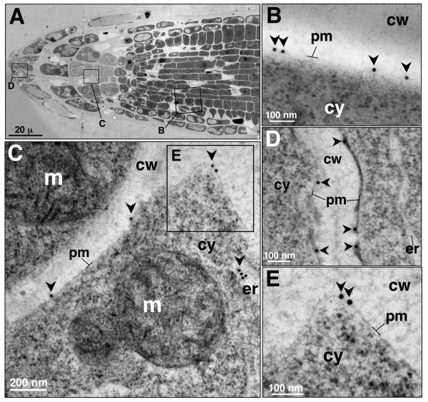
Figure 5.
Immunolocalization of AtCNGC10 to the plasma membrane of Arabidopsis root cells using TEM. (A) Transverse section of root tip showing three regions (rectangles) examined under TEM labeled with anti-AtCNGC10 antiserum. (B) Root meristematic cell plasma membrane; (C) Root columella cell; m, mitochondria; er, endoplasmic reticulum. (D) border tip cell. (E) Immunolabeled region in the box from panel C under higher magnification. 15 nm gold anti-rabbit secondary antibody used in all labeling. Cell wall, cw, and cytoplasm, cy. Size bars are in nanometers (nm).

