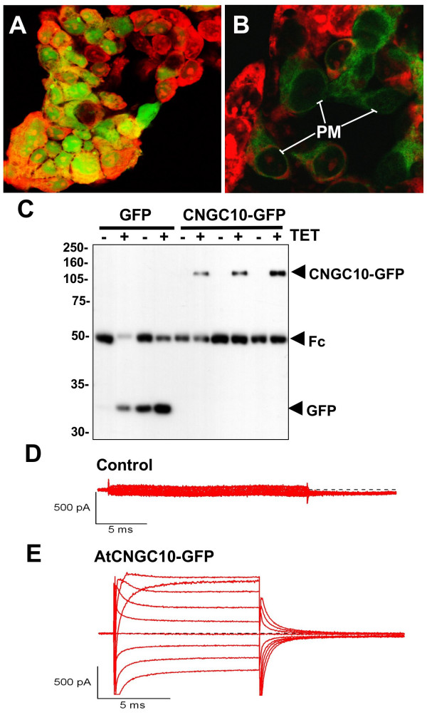
Figure 9.
Expression of AtCNGC10-GFP in HEK293 cells and patch clamp assays. Transient expression of (A) GFP alone in the vector and (B) the AtCNGC10-GFP fusion in HEK293 cells and visualized under the fluorescence microscope. HEK cells were fixed with 4% dimethyl-formamide and stained with propidium iodide (red fluorescence). Distinct GFP labeling of peripheral plasma membrane (PM) is marked. Immunoprecipitation of the cells expressing each construct with and without the inducer tetracycline (+/-) TET is shown. The bands corresponding to the GFP alone and AtCNGC10-GFP fusion are labeled. The Fc region of the antiserum used in immunoprecipitation is marked. Proteins were detected on immunoblots with the anti-FLAG antibody. ((D) Non-transfected HEK cell showed no measurable current. (E) HEK cell transfected with AtCNGC10-GFP showed large inward and outward currents in asymmetrical solution consisting of 145 K+out/145 NMDGin. Shown are leak-subtracted currents generated from command voltages between -60 to +100 mV in the presence of 100 μM dibutyryl cGMP.

