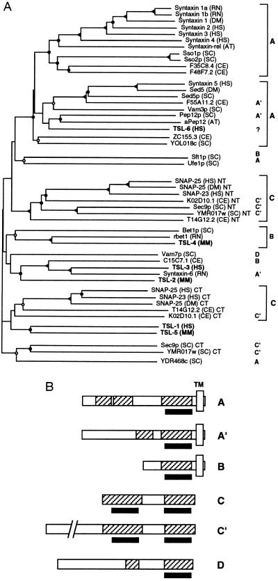
Figure 2.
(A) Nearest-neighbor dendrogram of the t-SNARE homology domain. The same set of sequences as in Fig. 1 is shown. Bifurcation points confirmed in 80–100 bootstrap replicates (out of 100) are marked by solid triangles, in 50–80 replicates are marked by solid circles, in 20–50 replicates are marked by open circles, and in less than 20 replicates are marked by no label. Boldface type on the right refers to the domain structures in B. Species abbreviations are as in Fig. 1. The newly identified SNARE homologues (TSLs) are indicated in boldface type. (B) Representative domain structures of t-SNARE superfamily members. Shaded boxes indicate coiled-coil regions. Open boxes labeled TM indicate putative transmembrane domains. The position of the t-SNARE coiled-coil homology domain is indicated by a solid bar at the bottom of the sequence representation. The chosen representative proteins are: A, syntaxin 1A; A′, Pep12p; B, Bet1p; C, SNAP-25; C′, Sec9; D, Vam7p.

