Abstract
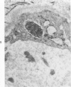
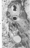
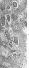
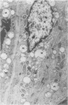
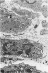
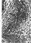
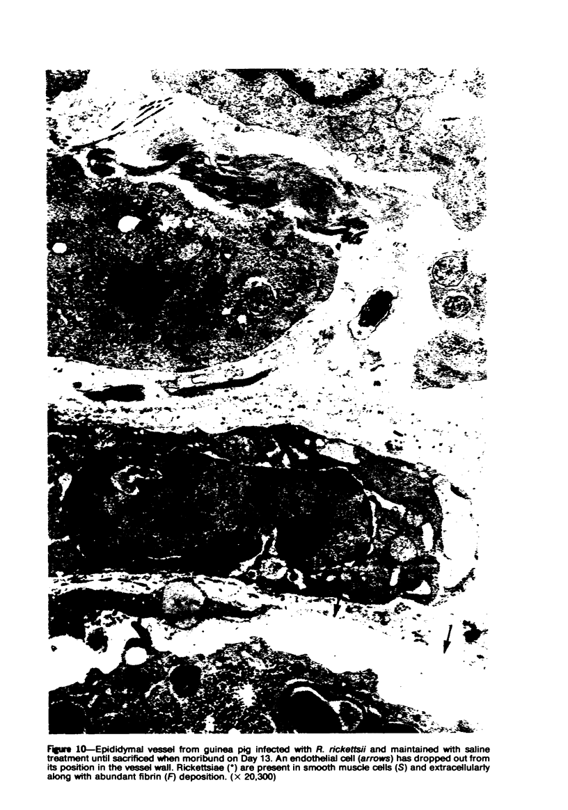
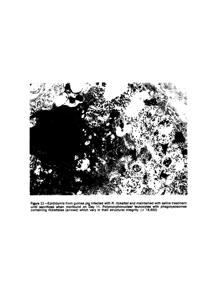
Moribund guinea pigs infected with Richettsia rickettsii were examined by necropsy, histology, immunofluorescence, electron microscopy, and serology. Untreated animals died at 9 and 10 days after inoculation. Animals given saline subcutaneously survived from 1 to 4 days longer. Prolonged survival was accompanied by more severe lesions: scrotal necrosis; infarction of ears; and swollen, hemorrhagic footpads, epididymis, and cremaster muscle. Histopathologic examination demonstrated that acute, necrotizing vasculitis, perivascular hemorrhage, and focal necrosis were more extensive. Direct immunofluorescence indicated many more rickettsiae in endothelium and vascular wall of saline recipients. Ultrastructurally, typical rickettsiae were present focally in the cytoplasm of endothelial and vascular smooth muscle cells. Cytopathology in infected and adjacent cells included swelling, mitochondrial enlargement with decrease in matrix density and loss of cristae, and increased pinocytosis. In addition, treated animals had more cytonecrosis, thrombosis, extravascular fibrin deposition, prominent inflammatory cells with polymorphonuclear phagocytosis of rickettsiae, and antibody production.
Full text
PDF
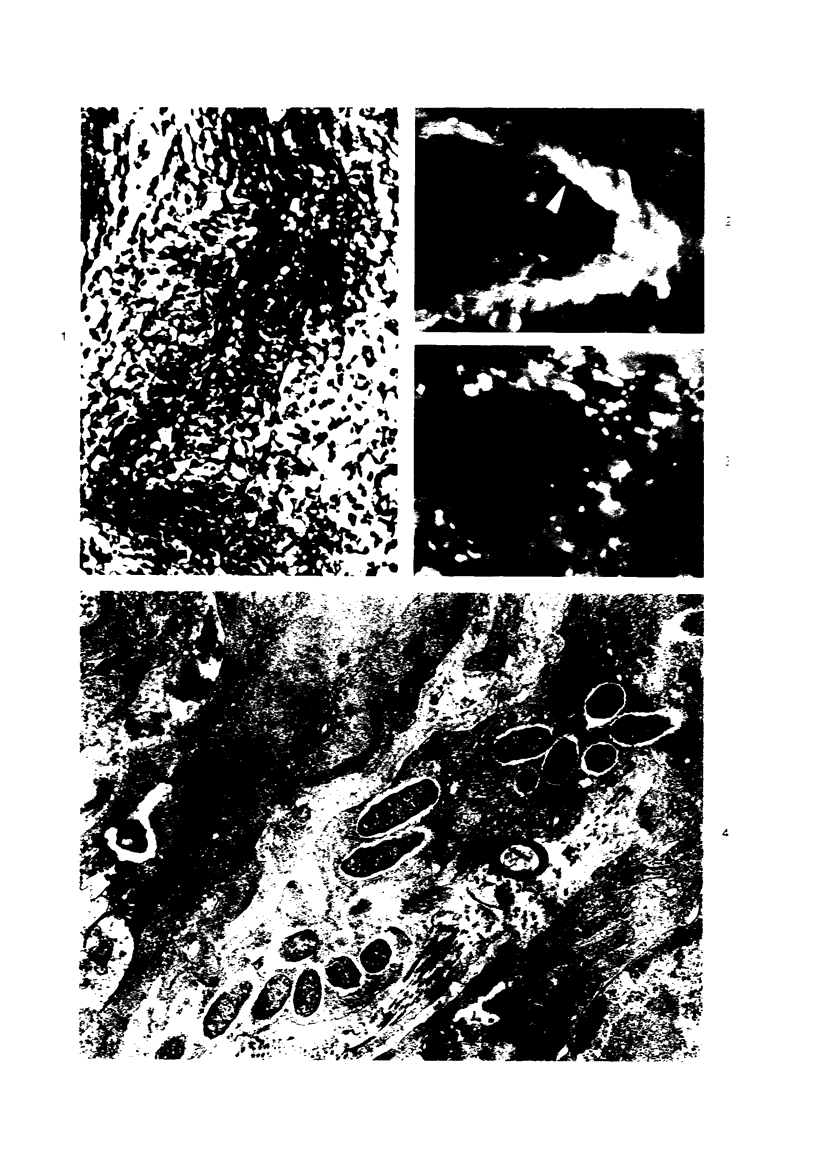
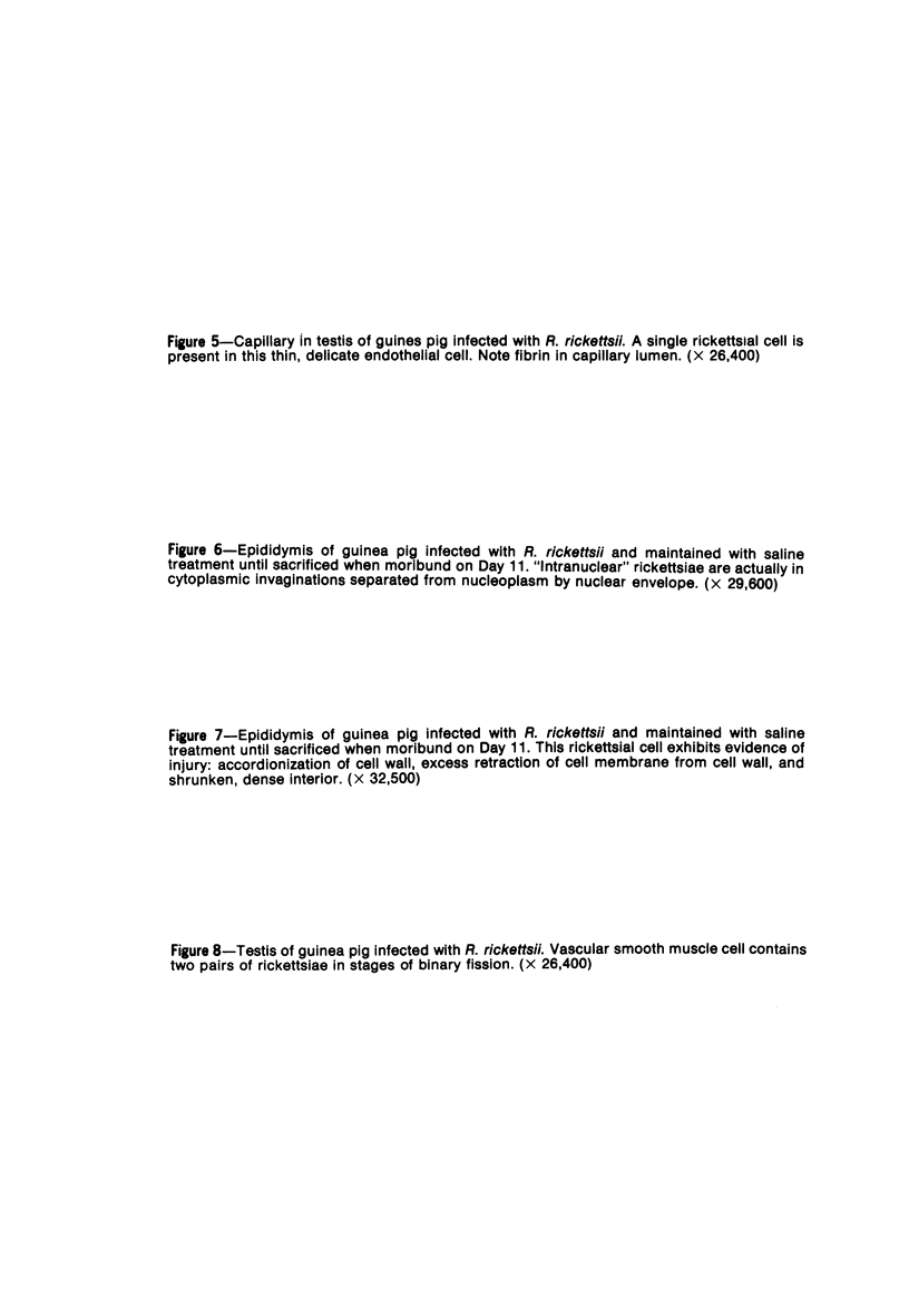
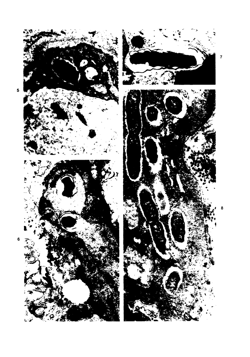
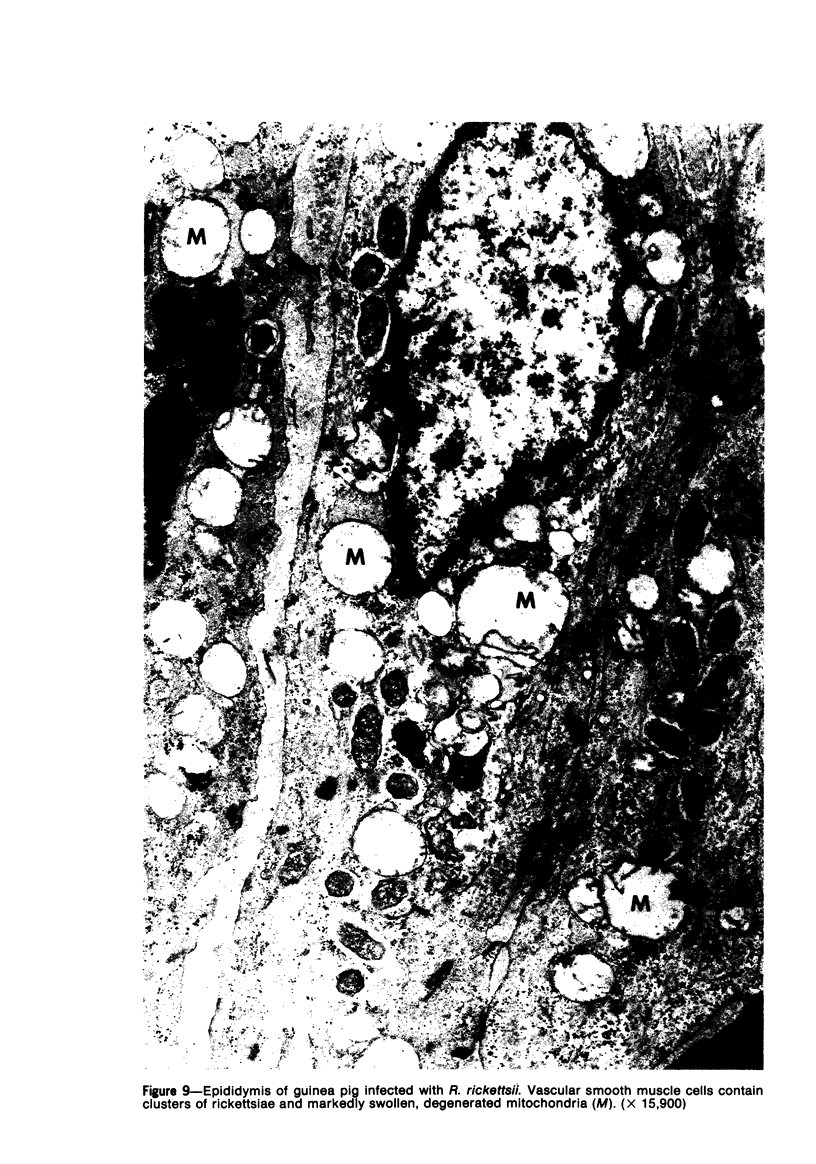
Images in this article
Selected References
These references are in PubMed. This may not be the complete list of references from this article.
- ALKAWA J. K., HARRELL G. T. Changes in the tissue radiosodium space associated with experimental Rocky Mountain spotted fever in guinea pigs. J Infect Dis. 1953 Nov-Dec;93(3):263–265. doi: 10.1093/infdis/93.3.263. [DOI] [PubMed] [Google Scholar]
- Allen A. C., Spitz S. A Comparative Study of the Pathology of Scrub Typhus (Tsutsugamushi Disease) and Other Rickettsial Diseases. Am J Pathol. 1945 Jul;21(4):603–681. [PMC free article] [PubMed] [Google Scholar]
- Fiset P., Ormsbee R. A., Silberman R., Peacock M., Spielman S. H. A microagglutination technique for detection and measurement of rickettsial antibodies. Acta Virol. 1969 Jan;13(1):60–66. [PubMed] [Google Scholar]
- MOLLENHAUER H. H. PLASTIC EMBEDDING MIXTURES FOR USE IN ELECTRON MICROSCOPY. Stain Technol. 1964 Mar;39:111–114. [PubMed] [Google Scholar]
- VENABLE J. H., COGGESHALL R. A SIMPLIFIED LEAD CITRATE STAIN FOR USE IN ELECTRON MICROSCOPY. J Cell Biol. 1965 May;25:407–408. doi: 10.1083/jcb.25.2.407. [DOI] [PMC free article] [PubMed] [Google Scholar]
- Weiss E. Growth and physiology of rickettsiae. Bacteriol Rev. 1973 Sep;37(3):259–283. doi: 10.1128/br.37.3.259-283.1973. [DOI] [PMC free article] [PubMed] [Google Scholar]
- de Brito T., Hoshino-Shimizu S., Pereira M. O., Rigolon N. The pathogenesis of the vascular lesions in experimental rickettsial disease of the guinea pig (Rocky Mountain Spotted fever group). A light, immunofluorescent and electron microscopic study. Virchows Arch A Pathol Pathol Anat. 1973;358(3):205–214. doi: 10.1007/BF00543228. [DOI] [PubMed] [Google Scholar]
- de Brito T., Tiriba A., Godoy C. V., Penna D. O., Jordão F. M. Glomerular response in human and experimental rickettsial disease (Rocky Mountain spotted fever group). A light and electron microscopy study. Pathol Microbiol (Basel) 1968;31(6):365–377. doi: 10.1159/000162038. [DOI] [PubMed] [Google Scholar]


