Figure 2.
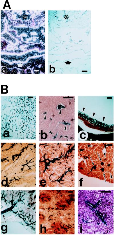
LacZ staining of an embryo at later embryonic stage (A) and in adult tissues (B). (Aa) In situ hybridization of embryos at E14.5 with LacZ probe. (b) Adjacent section of a, immunostained with anti-PECAM antibody. Identical staining pattern of a and b confirms specific and uniform endothelial expression of LacZ. ∗, Heart; arrow, dorsal aorta. (B) LacZ staining of adult brain (a, b), eye (c), heart (d), kidney medulla (e), kidney cortex (f), intestine (g), and spleen (h, i). In brain (a), virtually all vessels are stained. In higher magnification of the thinner sections of the brain (b), clearly demonstrates the EC staining (arrowheads). In eye (c), uniform and strong LacZ staining of the capillary plexus is indicated by arrowheads. In heart (d), uniform LacZ staining of the myocardial vasculature is evident. LacZ staining was also observed in endocardium (data not shown). In kidney cortex (f), all of the glomerular microvasculatures (arrows) are strongly LacZ-positive. Other microvasculatures are difficult to see with this low magnification unless they are clustered (arrowheads), but higher magnification confirmed the complete staining of all microvasculatures. In intestine (g), LacZ-positive mesenteric vasculature is indicated (arrow). In spleen, low magnification (h) and higher magnification (i) clearly demonstrates the uniform and EC-specific LacZ staining. (Bar = 100 μm.)

