Abstract
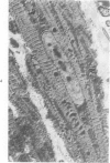
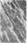
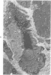
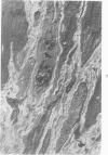
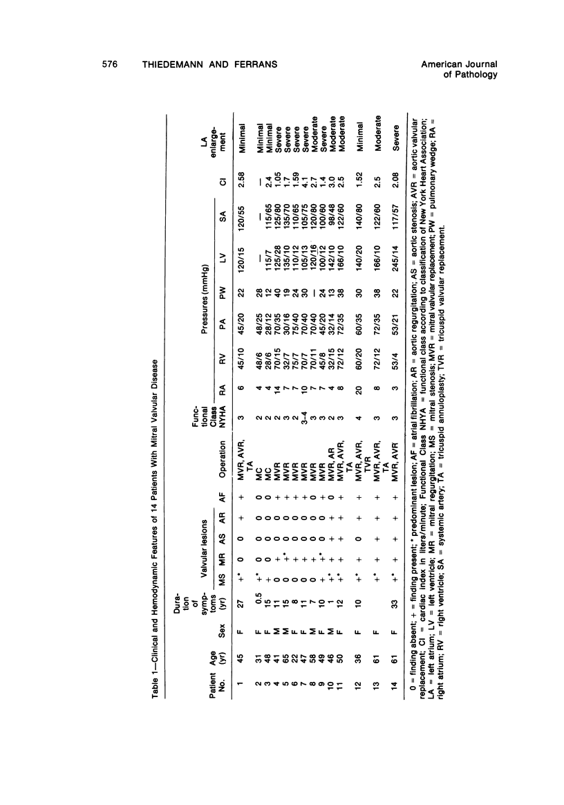
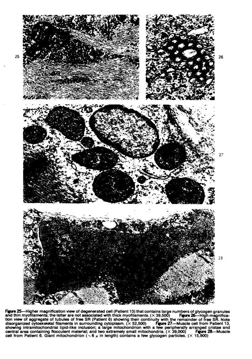
Light microscopic and ultrastructural observations were made on left atrial tissues obtained from 14 patients at the time of operation for correction of mitral valvular disease. Cardiac muscle cells varied in size but most frequently were hypertrophied. In fibrotic areas, present in all left atria, the muscle cells tended to be isolated from adjacent cells and exhibited degenerative changes of varying severity. These changes consisted or proliferation of Z-band material and cytoskeletal filaments, myofibrillar loss, proliferation of elements of free and extended junctional sarcoplasmic reticulum, variations in size and number of mitochondria, occurrence of abnormal mitochondria, dissociation of intercellular junctions, formation of spherical microparticles, and accumulation of lysosomal degradation products. Hypertrophy was considered to lead to cellular degeneration, with decrease or loss of contractile function. Atrial fibrillation was associated with severe cellular degeneration. The severity of degeneration was greater in patients with mitral regurgitation, with or without associated mitral stenosis, than in patients with pure mitral stenosis.
Full text
PDF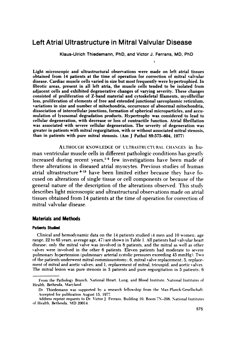




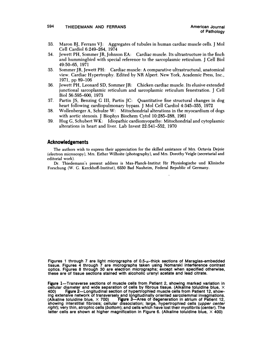
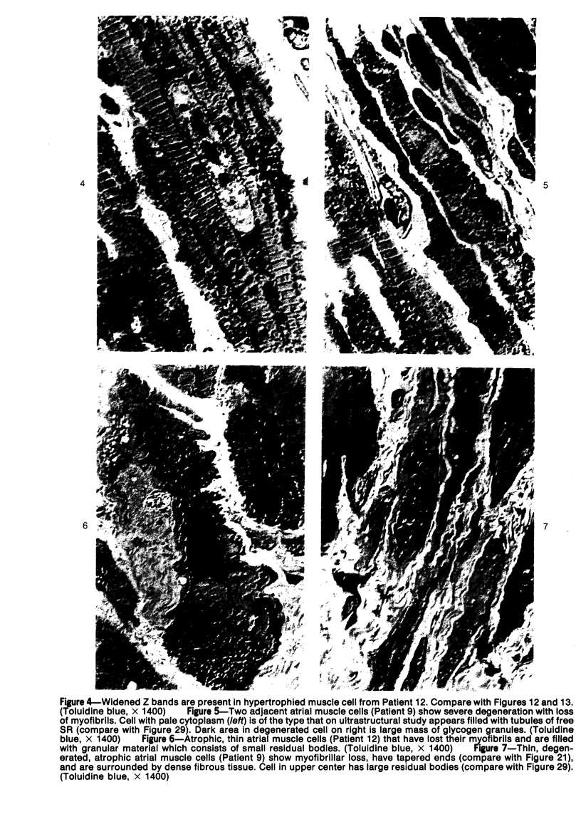
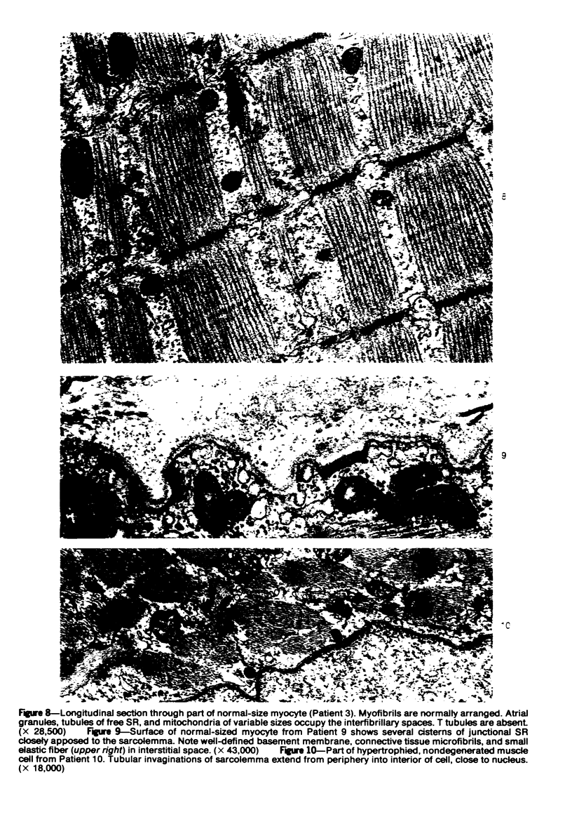
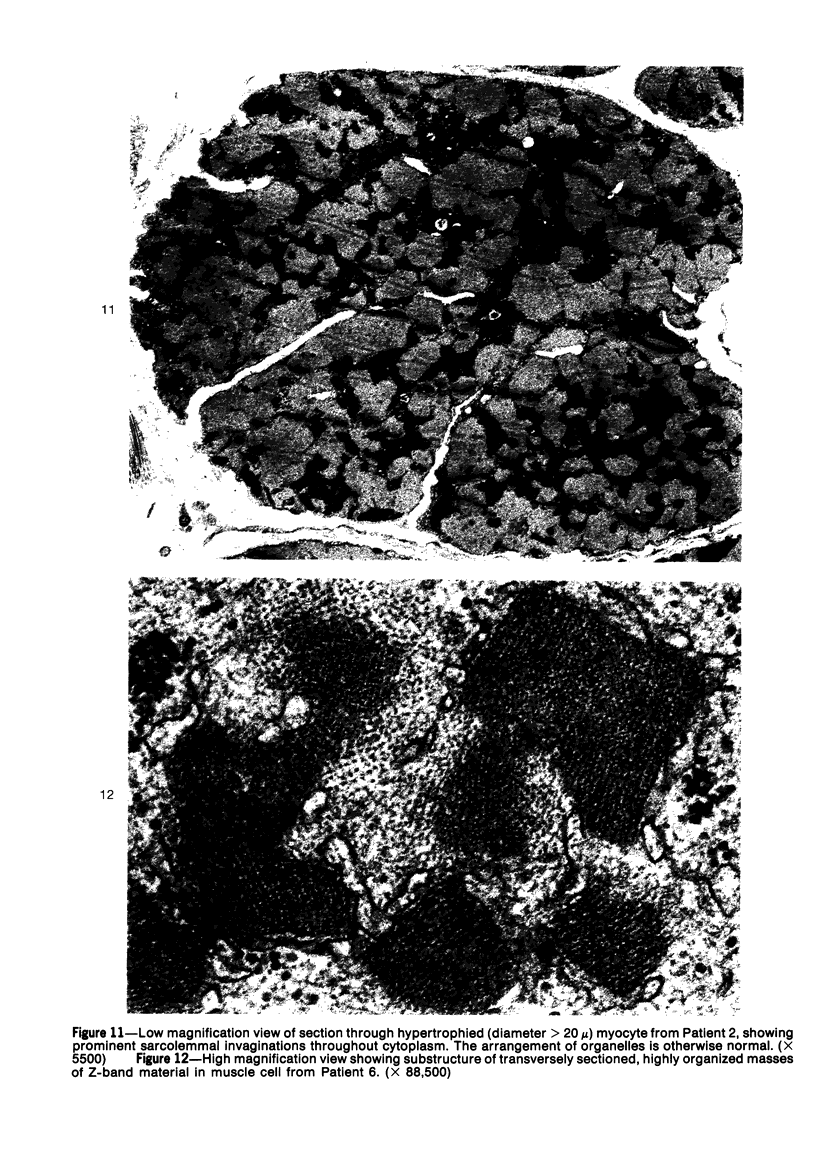

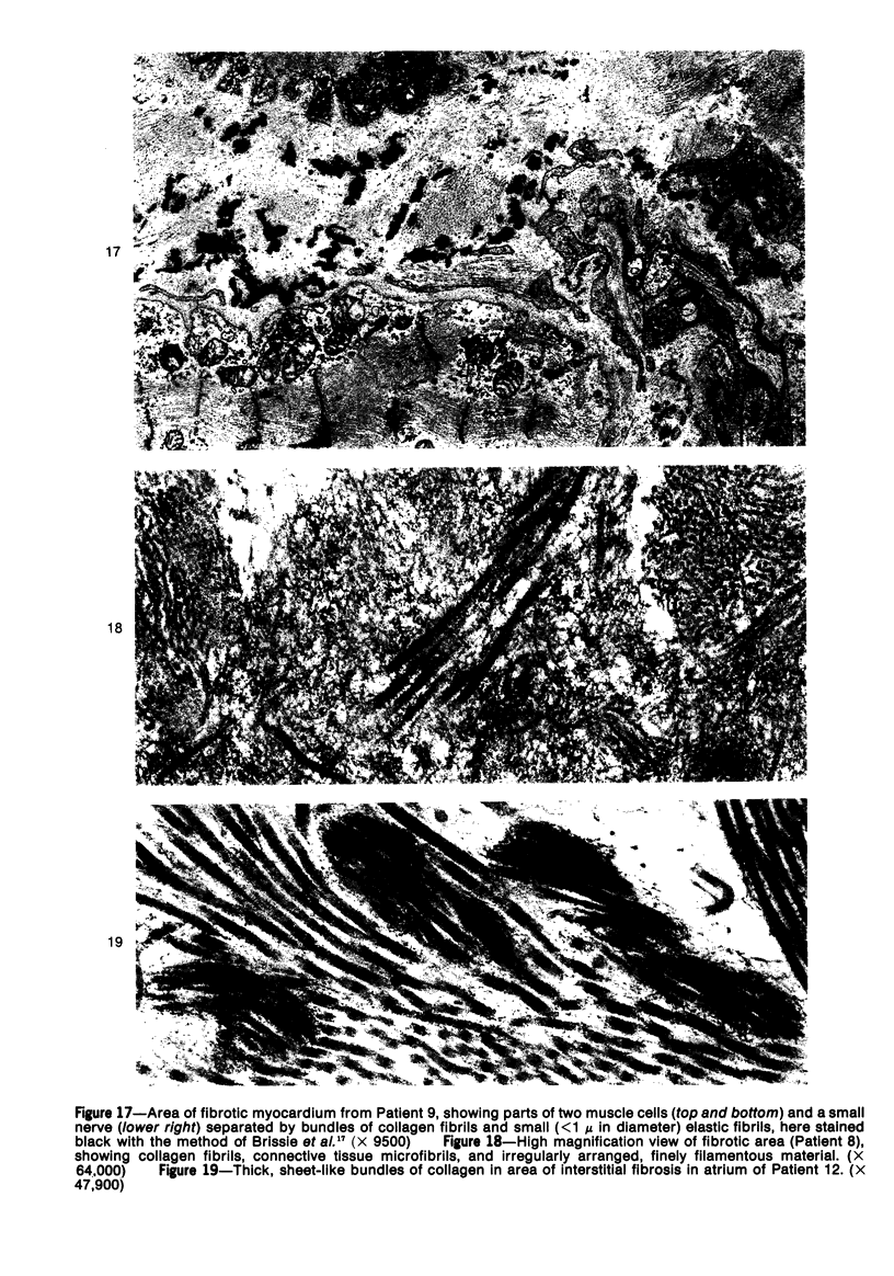

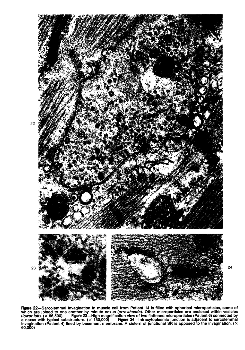

Images in this article
Selected References
These references are in PubMed. This may not be the complete list of references from this article.
- Ben-Ishay Z., Davies A. M., Laufer A. Fibrogenesis in the human myocardium: an electron microscopic study. Exp Mol Pathol. 1968 Jun;8(3):358–369. doi: 10.1016/s0014-4800(68)80005-x. [DOI] [PubMed] [Google Scholar]
- Bencosme S. A., Berger J. M. Specific granules in mammalian and non-mammalian vertebrate cardiocytes. Methods Achiev Exp Pathol. 1971;5:173–213. [PubMed] [Google Scholar]
- Bishop S. P., Cole C. R. Ultrastructural changes in the canine myocardium with right ventricular hypertrophy and congestive heart failure. Lab Invest. 1969 Mar;20(3):219–229. [PubMed] [Google Scholar]
- Brissie R. M., Spicer S. S., Hall B. J., Thompson N. T. Ultrastructural staining of thin sections with iron hematoxylin. J Histochem Cytochem. 1974 Sep;22(9):895–907. doi: 10.1177/22.9.895. [DOI] [PubMed] [Google Scholar]
- Buja L. M., Ferrans V. J., Levitsky S. Occurrence of intramitochondrial glycogen in canine myocardium after prolonged anoxic cardiac arrest. J Mol Cell Cardiol. 1972 Jun;4(3):237–254. doi: 10.1016/0022-2828(72)90061-2. [DOI] [PubMed] [Google Scholar]
- Buja L. M., Ferrans V. J., Maron B. J. Intracytoplasmic junctions in cardiac muscle cells. Am J Pathol. 1974 Mar;74(3):613–647. [PMC free article] [PubMed] [Google Scholar]
- Côté G., Mohiuddin S. M., Roy P. E. Occurrence of Z-band widening in human atrial cells. Exp Mol Pathol. 1970 Dec;13(3):307–318. doi: 10.1016/0014-4800(70)90093-6. [DOI] [PubMed] [Google Scholar]
- Fenoglio J. J., Jr, Wagner B. M. Studies in rheumatic fever. VI. Ultrastructure of chronic rheumatic heart disease. Am J Pathol. 1973 Dec;73(3):623–640. [PMC free article] [PubMed] [Google Scholar]
- Ferrans V. J., Massumi R. A., Shugoll G. I., Ali N., Roberts W. C. Ultrastructural studies of myocardial biopsies in 45 patients with obstructive or congestive cardiomyopathy. Recent Adv Stud Cardiac Struct Metab. 1973;2:231–272. [PubMed] [Google Scholar]
- Ferrans V. J., Thiedemann K. U., Maron B. J., Jones M., Roberts W. C. Spherical microparticles in human myocardium: an ultrastructural study. Lab Invest. 1976 Oct;35(4):349–368. [PubMed] [Google Scholar]
- Hug G., Schubert W. K. Idiopathic cardiomyopathy. Mitochondrial and cytoplasmic alterations in heart and liver. Lab Invest. 1970 Jun;22(6):541–552. [PubMed] [Google Scholar]
- Jewett P. H., Leonard S. D., Sommer J. R. Chicken cardiac muscle: its elusive extended junctional sarcoplasmic reticulum and sarcoplasmic reticulum fenestrations. J Cell Biol. 1973 Feb;56(2):595–600. doi: 10.1083/jcb.56.2.595. [DOI] [PMC free article] [PubMed] [Google Scholar]
- Jewett P. H., Sommer J. R., Johnson E. A. Cardiac muscle. Its ultrastructure in the finch and hummingbird with special reference to the sarcoplasmic reticulum. J Cell Biol. 1971 Apr;49(1):50–65. doi: 10.1083/jcb.49.1.50. [DOI] [PMC free article] [PubMed] [Google Scholar]
- Jones M., Ferrans V. J. Intramitochondrial glycogen in hypertrophied infundibular muscle of patients with congenital heart diseases. Am J Pathol. 1973 Jan;70(1):69–88. [PMC free article] [PubMed] [Google Scholar]
- Jones M., Ferrans V. J., Morrow A. G., Roberts W. C. Ultrastructure of crista supraventricularis muscle in patients with congenital heart diseases associated with right ventricular outflow tract obstruction. Circulation. 1975 Jan;51(1):39–67. doi: 10.1161/01.cir.51.1.39. [DOI] [PubMed] [Google Scholar]
- Lannigan R. A., Zaki S. A. Ultrastructure of the myocardium of the atrial appendage. Br Heart J. 1966 Nov;28(6):796–807. doi: 10.1136/hrt.28.6.796. [DOI] [PMC free article] [PubMed] [Google Scholar]
- Lannigan R. A., Zaki S. A. Ultrastructure of the normal atrial endocardium. Br Heart J. 1966 Nov;28(6):785–795. doi: 10.1136/hrt.28.6.785. [DOI] [PMC free article] [PubMed] [Google Scholar]
- Laufer A., Garces C. J., Ron N., Friedman I., Borman J. B. Morphological and immunological studies in valvular heart surgery. Pathol Microbiol (Basel) 1970;36(4):230–242. doi: 10.1159/000162452. [DOI] [PubMed] [Google Scholar]
- Legato M. J., Bull M. B., Ferrer M. I. Atrial ultrastructure in patients with fixed intra-atrial block. Chest. 1974 Mar;65(3):252–261. doi: 10.1378/chest.65.3.252. [DOI] [PubMed] [Google Scholar]
- Legato M. J. Sarcomerogenesis in human myocardium. J Mol Cell Cardiol. 1970 Dec;1(4):425–437. doi: 10.1016/0022-2828(70)90039-8. [DOI] [PubMed] [Google Scholar]
- Maron B. J., Ferrans V. J. Aggregates of tubules in human cardiac muscle cells. J Mol Cell Cardiol. 1974 Jun;6(3):249–264. doi: 10.1016/0022-2828(74)90054-6. [DOI] [PubMed] [Google Scholar]
- Maron B. J., Ferrans V. J. Intramitochondrial gylcogen deposits in hypertrophied human myocardium. J Mol Cell Cardiol. 1975 Sep;7(9):697–702. doi: 10.1016/0022-2828(75)90146-7. [DOI] [PubMed] [Google Scholar]
- Maron B. J., Ferrans V. J., Roberts W. C. Myocardial ultrastructure in patients with chronic aortic valve disease. Am J Cardiol. 1975 May;35(5):725–739. doi: 10.1016/0002-9149(75)90065-x. [DOI] [PubMed] [Google Scholar]
- Maron B. J., Ferrans V. J., Roberts W. C. Ultrastructural features of degenerated cardiac muscle cells in patients with cardiac hypertrophy. Am J Pathol. 1975 Jun;79(3):387–434. [PMC free article] [PubMed] [Google Scholar]
- McNutt N. S., Fawcett D. W. The ultrastructure of the cat myocardium. II. Atrial muscle. J Cell Biol. 1969 Jul;42(1):46–67. doi: 10.1083/jcb.42.1.46. [DOI] [PMC free article] [PubMed] [Google Scholar]
- Partin J. S., Benzing G., 3rd, Partin J. C. Quantitative fine structural changes in dog heart following cardiopulmonary bypass. J Mol Cell Cardiol. 1972 Aug;4(4):345–355. doi: 10.1016/0022-2828(72)90081-8. [DOI] [PubMed] [Google Scholar]
- Roy P. E., Dorais J., Morin P. J. Les foyers de nécrose intracelulaire focale dans le muscle cardiaque humain. Ann Anat Pathol (Paris) 1972 Jan-Mar;17(1):39–51. [PubMed] [Google Scholar]
- Roy P. E., Morin P. J. Dilatations of transverse tubules and of the intercalated disk in human cardiac muscle. J Mol Cell Cardiol. 1972 Aug;4(4):337–343. doi: 10.1016/0022-2828(72)90080-6. [DOI] [PubMed] [Google Scholar]
- Roy P. E., Morin P. J. Variations of the Z-band in human auricular appendages. Lab Invest. 1971 Nov;25(5):422–426. [PubMed] [Google Scholar]
- STEIN A. A., THIBODEAU F., STRANAHAN A. Electron microscopic studies of human myocardium. JAMA. 1962 Nov 3;182:537–540. doi: 10.1001/jama.1962.03050440029009. [DOI] [PubMed] [Google Scholar]
- Tomisawa M., Onouchi Z., Goto M., Nakata K., Kusunoki T., Asazuma S., Watanabe T., Hashimoto I. Congenital left atrial enlargement. A case report with special reference to myocardial fine structure. Jpn Circ J. 1975 Apr;39(4):417–424. doi: 10.1253/jcj.39.417. [DOI] [PubMed] [Google Scholar]
- WOLLENBERGER A., SCHULZE W. Mitochondrial alterations in the myocardium of dogs with aortic stenosis. J Biophys Biochem Cytol. 1961 Jun;10:285–288. doi: 10.1083/jcb.10.2.285. [DOI] [PMC free article] [PubMed] [Google Scholar]


