Figure 4.
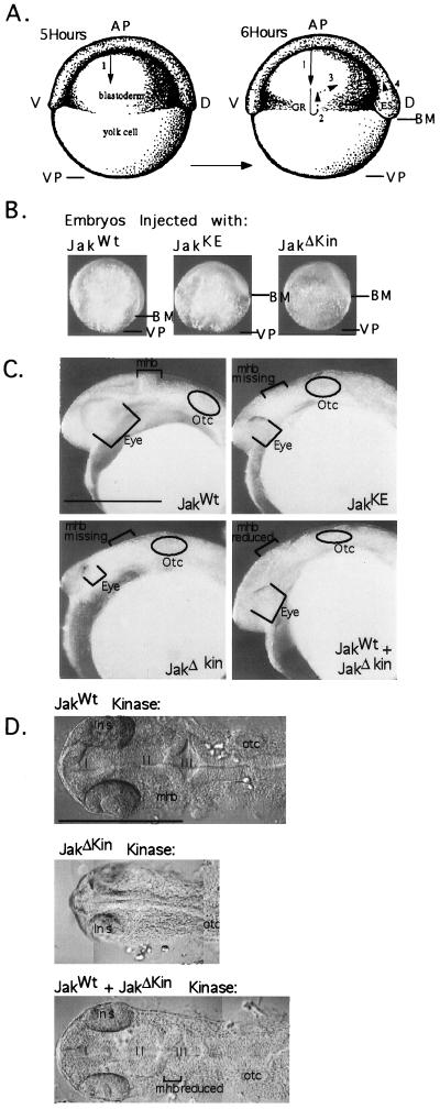
Defective Jak1 kinases inhibit cell movements of epiboly and reduce anterior structures. (A) During early zebrafish development four cell movements occur. Cells of the blastoderm move down the surface of the large yolk cell in a migration called epiboly (arrow 1). Once half of the yolk surface is covered, cells begin three additional cell movements. Involution (arrow 2) results in the formation of a localized thickening of cells known as the germ ring. Convergence of cells (arrow 3) toward the future dorsal side of the embryo creates the embryonic shield and extension (arrow 4) elongates the body axis. (B) One-cell embryos were injected with 100 pg of in vitro-synthesized RNA encoding JakWt, JakKE, and JakΔKin kinases. Until 4 h of development all embryos develop normally, however, at 4 h of development, when the cell migration of epiboly begins, embryos injected with defective forms of Jak1 kinase show impaired cell migration. At 8 h of development (shown above), embryos injected with JakWT have 80% of the yolk surface covered by blastomeres; in contrast, embryos injected with defective forms of Jak1 kinase have 50% of the yolk covered. The blastoderm margin, and hence the extent of cell migration, is indicated by the arrow marked BM. The vegetal pole (VP) is indicated. (C) By 24 h of development (lateral view), embryos injected with defective forms of Jak1 kinase exhibit reduced anterior structures; scale bar equals 0.5 mm. (D) In a dorsal view with Nomarski optics, these embryos possess small eyes, and neural structures anterior to the otocyst develop as a fused neural tube lacking ventricles and a midhind brain boundary. Injection of JakWT and JakΔKin gives partial rescue of anterior structures; scale bar equals 0.25 mm. AP, animal pole; VP, vegetal pole; V, ventral; D, dorsal; GR, germ ring; ES, embryonic shield; BM, blastoderm margin; Otc, otocyst; lns, lens; I, II, III, ventricles I, II, III; mhb, midhind brain boundary; scale bar equals 0.5 mm.

