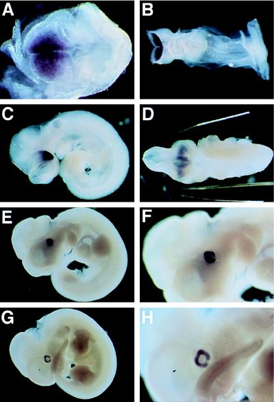
Figure 4.
Whole-mount in situ hybridization of E7.5 (A), E8.5 (B), E9.5 (C and D), E10.5 (E and F), and E11.5 mouse embryos (G and H). (Left) Anterior side of the embryos. (A) rax expression is detected in the cephalic head fold. (B) rax expression is at a high level in the prospective forebrain. (C) The embryo has finished turning and the optic vesicles have been formed. The optic vesicles exhibit a high level of expression of rax. (D) A ventral view of the embryo shown in C. rax expression is detected in the optic vesicles, optic stalk, and ventral diencephalon. (E–H) Lateral view of the embryos at E10.5 (E and F) and E11.5 (G and H). rax is expressed in the optic cup. F and H represent a higher magnification of E and G, respectively. Note that the optic fissures are negative for rax expression.

