Figure 6.
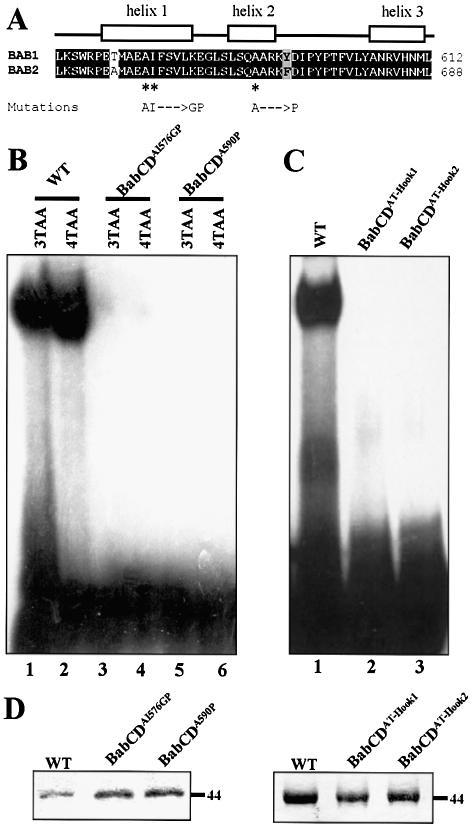
The AT-Hook and the psq motif are required for the DNA binding activity of the BabCD. (A) Indicated above the sequence alignment of the psq motif of Bab1 and Bab2 are the positions of three predicted alpha helices. The mutations made in helix 1 or in helix 2 of the psq domain of BabCD1 are shown below the sequences. (B) EMSAs with the 3TAA (lanes 1, 3 and 5) or the 4TAA (lanes 2, 4 and 6) oligonucleotides and the wild-type BabCD1 (lanes 1 and 2), or two psq domain mutants: BabCDAI576GP (lanes 2 and 4) and BabCDA590P (lanes 5 and 6). (C) EMSAs with the 4TAA oligonucleotide and the wild-type BabCD1 (lane 1), the BabCDAT-Hook1 (lane 2) or the BabCDAT-Hook2 (lane 3) mutants. (D) Coomassie blue-stained SDS–PAGE containing the indicated purified wild-type or mutant BabCD1 proteins. Their molecular mass is indicated in kDa on the right.

