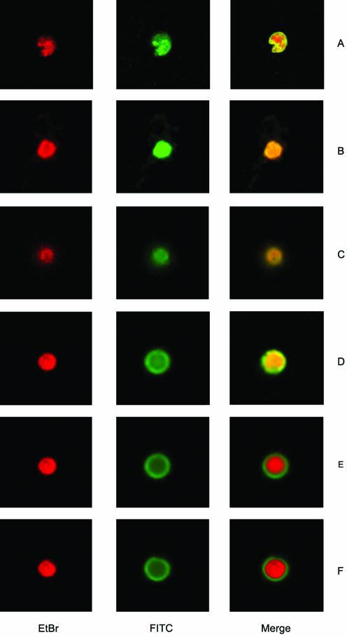
Figure 4.
Immunofluorescent localization of wild-type and C-terminally truncated mutant enzymes of LdTOP2 expressed in RS192. Yeast cells transformed with LdTOP2 wild-type and truncation constructs were prepared as spheroplasts, fixed and probed with anti-LdTOP2 antiserum. Visualization of the bound primary antibody was done with FITC- conjugated anti-rabbit IgG. Ethidium bromide staining was subsequently performed to highlight the nuclei and the area of the overlapping FITC and ethidium bromide stain are shown in the merged pictures. Cells were viewed at an original magnification of 100× under a Leica DM IRB inverted microscope. Images of cells expressing full-length L.donovani topoisomerase II (A), and mutants LdΔC1195 (B), LdΔC1118 (C), LdΔC1058 (D), LdΔC998 (E) and LdΔC785 (F) were captured and the fluorescent signal was photographed.

