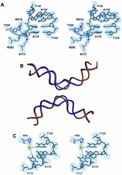
Figure 4.
(A) Stereoview of the 3Fobs–2Fcalc electron density map (contoured at 1.4 σ level) around the active site of the ‘cleavage-competent’ Cre subunit and the kink region of the loxP site. A metal ion (possibly a magnesium) is indicated by an orange sphere. (B) DNA backbone superposition of the wild-type loxP sequence as observed in the present structure (in blue) and the symmetrized loxS sequence (PDBID 4CRX; the second duplex was generated by applying 2-fold symmetry). While the overall bending is the same, marked differences are found in the spacer region resulting from different locations of kinks. (C) Stereoview of the 3Fobs–2Fcalc electron density map (contoured at 1.4 σ level) showing a magnesium-mediated protein–DNA contact.

