
Figure 5.
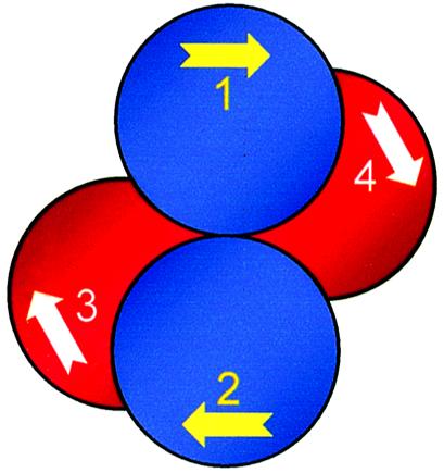
Model for BspMI. The subunits of the BspMI restriction endonuclease are illustrated as spheres, numbered 1–4. As with the NgoMIV tetramer (29), subunits 1 and 2 (in blue) are related by a two-fold axis and constitute a primary dimer, as do subunits 3 and 4 (in red). All four subunits contain a binding site for the asymmetric DNA sequence recognised by BspMI, in the direction marked by the arrow. The yellow arrows in subunits 1 and 2 indicate binding sites on the upper face of the protein and the white arrows in subunits 3 and 4 binding sites on the underside of the protein. When bound to subunits 1 and 2, the two recognition sequences are in antiparallel alignment. When bound to subunits 1 and 4, they lie parallel to each other.