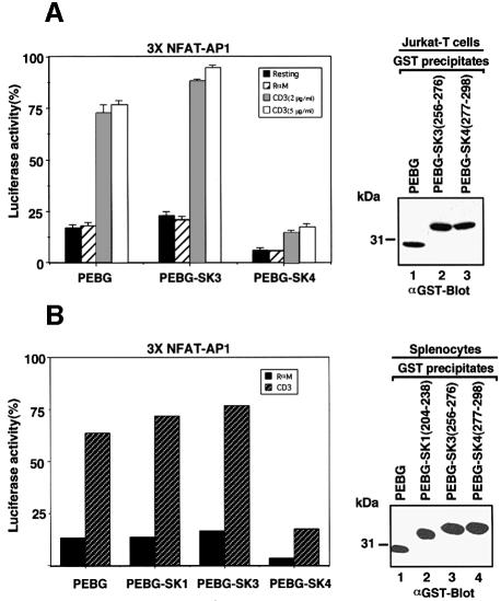
Fig. 6. SK4 peptide attentuates TcR up-regulation of IL-2 transcription. (A, left panel) Jurkat T cells were subjected to electroporation using 5 µg NF-AT of the IL-2 promoter luciferase reporter plasmid and 0.2 µg pRL-TK plasmid together with either 20 µg pEBG vector, pEBG-SK3 or pEBG-SK4 constructs. Cells were unstimulated (black bars) or exposed to rabbit anti-mouse (hatched bars), anti-CD3 (OKT3, 2 µg/ml, grey bars) or anti-CD3 (OKT3, 5 µg/ml, open white bars) for 6 h and assayed for luciferase activity. Luciferase units of the experimental vector were normalized to the level of the control vector in each sample. The data are representative of seven independent experiments. Results are the means and standard errors from three replicate experiments. (Right panel) Levels of GST fusion protein expression. Cell lysates from Jurkat T cells that had been transfected with GST–SK4 were subjected to immunoblotting with an anti-GST antibody. Lane 1, pEBG; lane 2, pEBG-SK3; lane 3, pEBG-SK4. (B, left panel) ConA-activated splenocytes 48 h after activation were subjected to electroporation using 5 µg NF-AT of the IL-2 promoter luciferase reporter plasmid and 0.2 µg pRL-TK plasmid together with either 20 µg pEBG vector, pEBG-SK1, pEBG-SK3 or pEBG-SK4 constructs. Cells were exposed to rabbit anti-mouse (black bars) or anti-CD3 (2C11, 2 µg/ml, hatched bars) for 6 h and assayed for luciferase activity. Luciferase activity was measured in spleen cells treated as described in Materials and methods. (Right panel) Levels of GST fusion protein expression. Cell lysates from spleen cells that had been transfected with GST–SK4 were subjected to immunoblotting with an anti-GST antibody. Lane 1, pEBG; lane 2, pEBG-SK1; lane 3, pEBG-SK3; lane 4, pEBG-SK4.

An official website of the United States government
Here's how you know
Official websites use .gov
A
.gov website belongs to an official
government organization in the United States.
Secure .gov websites use HTTPS
A lock (
) or https:// means you've safely
connected to the .gov website. Share sensitive
information only on official, secure websites.
