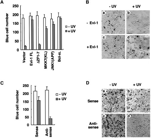
Fig. 6. Blocking of UV-induced cell death by Evi-1. (A) The 293 cells were transfected in duplicate with pSRα-βgal and the indicated plasmids. Then the cells were either left untreated or treated with 60 J/m2 UV, and stained for β-galactosidase expression. The number of blue cells in five randomly chosen fields was determined, and the data shown are averages of three separate experiments. (B) Colorimetric staining of vector (pME18S)- (a and b) or pME18S-Evi-1- (c and d) transfected cells either left untreated (a and c) or treated with UV (b and d). Scale bar = 150 µm. (C) HEC1B cells were treated with 5 µg of the sense or antisense oligonucleotide for Evi-1. Then the cells were either left untreated or treated with 100 J/m2 UV, and stained for β-galactosidase expression. The number of blue cells in five randomly chosen fields was determined, and the data shown are averages of three separate experiments. (D) Colorimetric staining of the sense (a and b) or antisense (c and d) oligonucleotide-transfected cells either left untreated (a and c) or treated with UV (b and d). Scale bar = 150 µm.

An official website of the United States government
Here's how you know
Official websites use .gov
A
.gov website belongs to an official
government organization in the United States.
Secure .gov websites use HTTPS
A lock (
) or https:// means you've safely
connected to the .gov website. Share sensitive
information only on official, secure websites.
